Anatomy of LUNGS and MCQs for NEET, GPAT,Nursing Exams
LUNGS
- These are the two cone shaped organs present in the thoracic cavity which are separated by other structures present in the mediastinum. Even if one lung collapses due to some trauma the other lung remain expanded.
- The lungs are protected by double layered wall called the pleural membrane, it is a serous membrane.
- The superficial layer called the parietal pleura lines the cavity while the deep layer to it called the visceral pleura covers each lung and in between these layer is a lubricating fluid called the pleural cavity.
- This fluid reduces friction and also allows the membrane to slide easily during the time of breathing.
- The fluid is secreted by the epithelial cells of the membrane .If either of the two membranes stops working due to some serious issues and the air is sucked it the pleural space, it results in collapsing of either the part of the lung or even the entire lung.
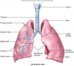
- Lungs have a base, apex coastal surface and medial surface.
- The first is the inferior portion of lungs called the base which is concave shaped and fits in the convex surface of diaphragm. At the superior end is a narrow edge called the apex.
- The coastal surface is the outer surface of the lung which lies against the cartilages , ribs and intercoastal muscles.
- The medial surface Of each lung as the name suggest, faces the other lung across the mediastinum.

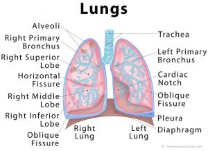
- One or two fissures divide the lung in lobes.
- The right lung is divided in 3 lobes while the left lungs contains 2 lobes, the left lung is smaller in size because the heart occupies the space left to the midline.
- The right lung has superior middle and inferior lobe.
- The oblique fissure separates the middle lobe from inferior lobe while the superior part of the oblique fissure separates the superior lobe from inferior lobe and the horizontal fissure separates the superior lobe from middle lobe, on the other hand the oblique fissure separates the superior and inferior lobe of the left lung.

- Each lobe receives the secondary (lobar) bronchi and within the lung the sec.
- Bronchi give rise to tertiary(segmental) bronchi.
- The segment of lung tissue where the tertiary bronchi supplies is known as bronchopulmonary segment, each bronchopulmonary segment of the lungs contains several small compartments called the lobules.
- The terminal bronchi divides in the smaller microscopic respiratory bronchi which again subdivides into several alveolar ducts. From trachea to the alveolar ducts there is presence of 25 orders of branching.
MULTIPLE CHOISE QUESTIONS(MCQs)
1. Where are the lungs located?
A. Inferior to the trachea B. Mediastinum of thoracic cavity
c. anterior to the esophagus D. Abdominal region
2. The pleural membrane is of?
A. serous membrane B. Mucous membrane
c. cuboidal epithelium D. Simple epithelium
3. how many lobes are present in right lung?
A. 3 B. 2
C. 4 D. 5
4. The oblique fissure of right lung separates which of the two lobes?
A. middle lobe from inferior lobe b. inferior lobe to superior
c. superior to inferior D. inferior lobe to the part of superior lobe
5. Which of the following statement is true?
A. The coastal surface lies directly across the mediastinum
B. The division between the lobes are known as lobules
C. The right lung contains the concavity k/as cardiac notch
D. Hilum is the region through which bronchi blood vessels lymphatic vessels enter and exit.
6. Which of the following is the key function of pleural cavity?
A. Reduces friction between membranes B. Slide easily on one another
c. allows membrane to adhere on one another D. all of the above
7. Match the following:-
(a) parietal cavity 1. Horizontal fissure
(b) visceral cavity 2. Lines the walls of thoracic cavity
(c) separates middle and superior lobe 3. Oblique fissure
(d) separates superior and inferior lobe 4. Covers the lungs
8. The respiratory bronchi subdivides in which of the following structure
A. Primary bronchi B. Lobar bronchi
C. Segmental bronchi D. none of the above
9. Why is the right lung is divided in 3 lobes while the left lung has 2 lobes?
a. It contains more space than left lung B. because right side is always better
C. right side contains cardiac notch D. all of the above
10. Inflammation of pleural cavity is called as
a. pleurisy B. pleural effusion
C. atelectasis D. Pneumothorax
ANSWERS:-
1. mediastinum of thoracic cavity
2. serous membrane
3. 3
4. inferior lobe from the part of superior lobe
5. hilum is the region through which bronchi blood vessels lymphatic vessels enter and exit
6. all of the above
7. (a) – 2 (b) – 4 (c) – 1 (d) – 3
8. none of the above
9. it contains more space than left lung
10. pleurisy
REFRENCES:-1. Ross and Wilson-Anatomy and physiology in health and illness; 12th edition; page no.-:250-251 .
2. Gerard J. Tortora -Principles of anatomy and physiology; edition twelfth ; page no.-: 885-887.
Participate in Online FREE GPAT TEST: CLICK HERE