Anatomy of Skin, Layer of Epidermis and Dermis and Important Questions for Exam
Skin or the integumetry system is a tough outer protective layer covering the body organs. Skin itself is known as a organ because it consist of different types of tissues that are joint together to perform some specific functions. It is the largest organ of the body in surface area (20 sq feet) and weight(4.5-5 kg).
ANATOMY
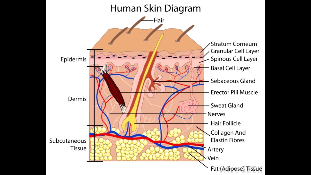
The skin consist of 3 layers, the superficial thinner layer called the epidermis and the inferior thicker layer called the DREMIS. The epidermis is made up of epithelial tissue nad the dermis is of connective tissue, just below the dermis is the subcutaneous layer called the hypodermis which is made up of areolar and adipose tissue.
EPIDERMIS:-
The epidermis is composed of keratinized stratified squamous epithelium. It contains 4 types of cells
• Keratinocytes – arranged in 3-4 layers and contain keratin which protects the tissues from heat and chemicals. These cells also produce lamellar granules which prevents entry of water into the skin.
• Merkel cells – these are least in number and are present in the deepest layer of epidermis, their function is to detect touch sensations.
• Langerhans cells – these arise from red bone marrow and are then transported to epidermis, they work for immune responses against microbes, they also activate other cells to perform immune surveillance.
• Melanocytes – these are 8% of epidermal cells and produce a pigment called melanin which is responsible for skin color and absorbs the UV light.
The epidermis majorly have 5 layers.
1. Stratum Basale – the deepest layer containing cuboidal and columnar keratinocytes . some cells in this layer are also known as stem cells as they produce different cells. Melanocytes and Merkel cells also found here. This layer is also known as stratum germinativum.
2. Stratum Spinosum – superior to Basale and has 8-10 layers of many sided keratinocytes which are closely packed, this type of arrangement provide strength and flexibility to the skin, Langerhans cells are present here.
3. Stratum Granulosum – this layer is in middle of the epidermis and consist of 3- 4 layers of flattened keratinocytes which undergoes apoptosis. They also contain lamellar granules that prevent water entry, this layers contains dead cells.
4. Stratum lucidum – it contains 3-5 layers of clear flattened dead keratinocytes.
5. Stratum Corneum – this layer contain 25-30 layers of dead flattened keratinocytes , contains keratin and lamellar granules in large amount.
In our body there are two types of skin present
1. Thick skin – it contains all the five layers of epidermis are present in areas where exposure to friction is higher like in palms soles fingertips etc.
2. Thin skin – it contains 4 layers of epidermis, stratum lucidum is absent in such areas where exposure to friction is less.
Dermis:-
This is the deeper layer of the skin which is made up of connective tissue consist of collagen elastic fibers because of which dermis has great tensile strength and also has the ability to stretch and recoil easily. It contains different type of cells like fibroblast macrophages adipocytes etc. Blood supply to the epidermis is also done by dermis. Dermis mainly have 2 layers:-
• The papillary region- superior portion of dermis which almost covers one-fifth part of dermis, consist of areolar connective tissue, contains dermal ridges Meissner corpuscles and nerves.
• The reticular region- the deeper portion of dermis and covers 4-5th of the dermis and contains irregular connective tissue with collagen and elastic fibers. Space between fibers contains cells hair follicles sebaceous glands and sudoriferous glands.
MULTIPLE CHOICE QUESTIONS (MCQs)
1. Which of the following epidermal layer is not found in thin skin?
A. Stratum Corneum B. Stratum lucidum
C. Stratum spinosum D. Stratum granulosum
2. Which of the following is not the function of skin?
A. calcium production B. PROTECTION
C. excretion of wastes D. temp regulation
3. Which of the following layer is also known as stratum germinativum?
A. stratum corneum B. stratum Basale
c. stratum spinosum D. stratum lucidum
4. Which of the following cells are responsible for our skin color?
A. melanocytes B. keratinocytes
c. Markel cells D. Langerhans cells
5.Which of the following statement is not true?
A. Keratinocytes protects skin and muscles from heat and chemicals
B. papillary region is responsible for the figure prints
C. Markel cells are used for touch sensations
D. Apoptosis takes place in stratum granulosum
6. Which of the following is the feature of reticular region?
A. hair follicles B. epidermal ridges
C. Sebaceous glands D. all of the above
7. What is the function of Meissner corpuscles present in the papillary region?
A. touch sensations B. Sensation of warmth
C. sensation of coolness D. pain sensations
8. Match the following
A. prevents water entry 1. Langerhans cells
B. sensitive to pressure 2.Sebacous glands
C. works for immune system 3. Pacinian corpuscles
D. protect the skin hairs from drying
And becoming brittle 4. Lamellar granules
9. What is the main function of Pacinian corpuscles present in the hypodermis?
A. tickling sensations B. Itching sensation
C. blood supply to the hypodermis D. none of the above
10. What is abnormal thickening of stratum Corneum called?
A. Dandruff B. callus
C. Comedo D. Abrasion
ANSWERS:-
1. stratum lucidum
2. calcium production
3. stratum Basale
4. Melanocytes
5. papillary region is responsible for finger prints
6. all of the above
7. touch sensations
8. a – 4 b – 3 c – 1 d – 2
9. none of the above
10 callus
Participate in Online FREE GPAT TEST: CLICK HERE
Participate in Online FREE Pharmacist TEST: CLICK HERE
Participate in Online FREE Drug Inspector TEST: CLICK HERE
REFERENCES:-
1. Ross and Wilson-Anatomy and physiology in health and illness; 12th edition; page no.-:362-363.
2. Gerard J. Tortora -Principles of anatomy and physiology; edition twelfth ; page no.-:148-153 .