Introduction to Central Nervous System Anatomy and MCQs for NEET, GPAT and Competitive Exams
CENTRAL NERVOUS SYSTEM
The CNS is divided in two essential pats: the brain and spinal cord. Both these parts are surrounded and protected; brain is enclosed by the skull and the spinal cord is protected by vertebral column. The membranous covering known as meninges is also found between the skull and brain, and between spinal cord and vertebral column for protection.
Meninges
The brain and spinal cord are completely surrounded and protected by three layer of tissue; these are
- DURA MATER :- dura mater in brain consist of two layers of tissue. The outer layer takes place of periostenum of skull bones and the inner layer provides complete covering of the brain. There is only a potential space between the two two layers; except where the inner layer sweep inwards between the cerebral hemispheres of cerebrum to form falx cerebri, between cerebellar hemispheres to form falx cerebella.
Spinal dura mater forms a loose sheath around the spinal cord. It extends from foramen magnum to the S2. It is extension of the inner layer brain’s dura mater. it is separated by the periostenum of the vertebral column and ligaments of neural cavity by the epidural space. The epidural space contains blood vessels and areolar connective tissue.
- ARACHNOID MATER:- It is a layer of fibrous tissue that lies between the dura mater and the pia mater. It is separated from the dura mater by the subdural space that contains large amount of serous fluid; it is separated from the pia mater by the subarachnoid space, which contains cerebrospinal fluid.
- PIA MATER:– This is a delicate layer of connective tissue which contains many small blood vessels.
Ventricles of brain
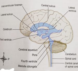
The brain contains four irregular shaped cavities also known as ventricles; these are:- left and right lateral ventricle, third ventricle and fourth ventricle.
- LATERAL VENTRICLE:- these ventricles lies between the cerebral hemispheres, one on each side of median plane. And just below the corpus callosum. Septum lucidum is a thin membrane that separate the two lateral ventricles. They communicate with the third ventricle through the interventricular foramina.
- THIRD VENTRICLE:– it is situated below the lateral ventricle, and between the two parts of the thalamus. It communicates with the fourth ventricle by a canal known as cerebral aqueduct.
- FOURTH VENTRICLE:– it is a diamond-shaped cavity which lies below and behind the third ventricle; between the cerebellum and pons. It is continous with the central canal of the spinal cord and communicates with the subarachnoid space by the foramina in its roof.
Cerebrospinal fluid(CSF)
The CSF is a clear, colorless liquid that protects the brain and spinal cord from physical and chemical injuries. CSF continuously circulates through the cavities of brain and spinal cord and also circulates around the brain and spinal cord in the subarachnoid space, between the arachnoid mater and pia mater. The main constituent of CSF are-
- Glucose
- Proteins
- Urea
- Lactic acid
- Cations
- Anions
- And some WBCs
Formation of CSF
The main site for the production of CSF is the choroid plexuses. Choroid plexuses are the network of capillaries which lies in the wall of the ventricles. These plexuses are surrounded or covered by the ependymal cells. These cells forms CSF from blood plasma through filtration and secretion. These cells acts as blood-cerebrospinal fluid barrier, hence prevents leakage or backflow of the fluid.
Circulation of CSF
The CSF formed by the choroid plexuses of each lateral ventricle, then flows into the third ventricle through the interventricular foramina. More CSF is added in the third ventricle through its own choroid plexuses; the total fluid then passes into the fourth ventricle through the aqueduct of midbrain. Similarly, the choroid plexuses of 4th ventricle also contribute some fluid. This fluid then enters the subarachnoid space through 3 openings: median aperture and two lateral aperture located in the roof of the 4th ventricle. CSF then circulates in the central canal of spinal cord and around the subarachnoid space. CSF is reabsorbed slowly by the arachnoid villi of arachnoid mater. Through these valli, the CSF returns back to the venous sinuses.
Functions of CSF
The three main functions of CSF include-
- Mechanical protection:- CSF acts as a shock-absorbing medium, which protects the delicate tissues of brain and spinal cord from jolts.
- Chemical protection:- CSF provides a needed chemical environment for the transmission of the signals.
- Circulation:- CSF acts as a transporter, and allows exchange of nutrients and waste products between the blood and the nervous tissue.

Multiple choice questions(MCQs)
1. Which of the following is not the part of CNS?
A. Brain B. grey matter
C. spinal cord D. both A and C
2. Which cells encloses the choroid plexuses?
A. ependymal cells B. astrocytes
C. oligodendrocytes D. microglia
3. what is the function of CSF?
A. mechanical protection B. chemical protection
C. circulation D. all of the above
4. from where does the CSF mixes with the venous drain?
A. arachnoid villi B. central canal
C. lateral column of grey matter D. none of the above
5. match the following-
a. dura mater 1. Lies between two parts of thalamus
B. pia mater 2. Lies between cerebellum and pons
c. fourth ventricle 3. Layer of delicate tissue
d. third ventricle 4. Consist of 2 layers of tissue
6. In which ventricle, the openings like median aperture and lateral aperture lies?
A. third ventricle B. right lateral ventricle
C. left lateral ventricle D. fourth ventricle
7. Which of the following Is not the constituent of the cerebrospinal fluid?
A. glucose B. proteins
C. lactic acid D. none of the above
8. Which of the following statement is NOT true?
A. meninges lies between only brain and skull
B. Third ventricle lies between two parts of thalamus
C. fourth ventricle lies below and behind 3rd ventricle
D. lateral ventricles contain median and aperture openings
9. What separates the dura mater from the arachnoid mater?
A. arachnoid space B. arachnoid villi
C. subdural space D. periostenum
10. Where does the meninges of brain lies?
A. between skull and brain B. around the skull
C. between spinal cord and vertebral column D. both A and C
ANSWERS:-
1. both A and C
2. ependymal cells
3. all of the above
4. arachnoid villi
5. a – 4 b – 3 c – 1 d – 2
6. Fourth ventricle
7. none of the above
8. meninges lies between only brain and skull
9. none of the above
10. both A and C
Participate in Online FREE GPAT TEST: CLICK HERE
Participate in Online FREE Pharmacist TEST: CLICK HERE
Participate in Online FREE Drug Inspector TEST: CLICK HERE
REFERENCE: 1. Ross and Wilson-Anatomy and physiology in health and illness; 12th edition; page no.-: 152-154.