Parts of BRAIN: CEREBRUM Anatomy and MCQs For NEET, GPAT, Staff Nurse, Lab Technician and SSC Exam
BRAIN
The brain is a large organ which lies in the cranial cavity. The average weight of brain is 1.4 kg. It is divided in 4 parts: cerebrum, brain stem, diencephalon and cerebellum.
CEREBRUM
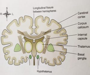
Cerebrum is the largest part of the brain. It lies in the anterior and middle cranial fossae. Cerebrum is divided by a deep cleft known as longitudinal cerebral fissure into right and left cerebral hemispheres. Each of the hemisphere contains one lateral ventricle. Deep inside the brain, these hemispheres are connected by a white matter mass known as corpus callosum. Falx cerebri is formed by the dura mater ; separates two hemispheres and which leads to the corpus callosum.
The superficial part of cerebrum is composed of cell bodies and is known as cerebral cortex. Deep layers consist of nerve fibers and white matter. The cerebral cortex contains various infoldings and furrows. The found areas of the folds are known as gyri(convolutions). These gyri are separated by sulcus(fissures). The gyri increases the surface area of the cerebrum.
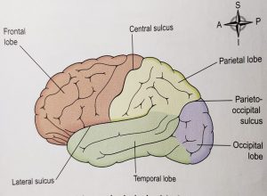
Each hemisphere of cerebrum is divided in different lobes. These lobes are named according to the bone where they lie; like: frontal, parietal, occipital and temporal. the boundries of these lobes are made by the fissures and are named as:- central, lateral and parieto-occipital sulcus.

CEREBRAL TRACTS:- Within the cerebrum, the lobes are connected by the mass of nerve fibers known as cerebral tracts. These tracts makes the white matter of the brain. The afferent and efferent fibers (tracts) which links the different part of brain are:-
- association tracts:- numerous in number, connects different parts of cerebral hemispheres
- Commissural tracts:- connects the corresponding areas of the two cerebral hemispheres.
- Projection tracts:- connects the cerebral cortex with the grey matter of lower parts of the brain and with spinal cord. for eg- internal capsule.
The internal capsule is an important projection tract which lies within the brain between the basal ganglia and the thalamus. The fibers that form the internal capsule carries many nerve impulses to and from the cerebral cortex. Motor fibers within the capsule form the pyramidal tracts which crosses the medulla oblongata and are main pathway for skeletal muscles.

BASAL GANGLIA:- these are the group of cell bodies that lie within the brain and are the pat of extrapyramidal tract . These are the relay stations having connection to many parts of the brain. Their main functions include: initiation and control of complex movements and coordinated activities.
Functions of cerebral cortex:- the three main types of activities associated with cerebral cortex include
- higher order functions: mental activities involved in memory, sense of responsibility, thinking reasoning etc.
- sensory perceptions: includes perception of pain, temp, touch, sight , hearing etc.
- initiation and control of skeletal muscle contraction and voluntary movement.
Functional areas of cerebral cortex:- the main functional areas of cerebral cortex are involved exclusively only in on function each. The areas of cortex which lies anterior to the central sulcus are involved in motor functions; an those lying posterior to the central sulcus are associated with sensory functions.
- Motor areas of cerebral cortex are as follows:-
1. Primary motor area:– it lies in the frontal lobe anterior to the central sulcus. they control the skeletal muscle activity.
2. Motor speech (broca’s) area:– it lies in the frontal sulcus, above the lateral sulcus. It controls muscle movements for speech.
- Sensory areas of cerebral cortex are as follows:-
1. Somatosensory area:– it lies behind the central sulcus. These areas receives sensations of pain, touch, awareness of muscular movement and position of joints.
2. Auditory area:– lies below the lateral sulcus within the temporal lobe. This area receives and interpret the signals transmitted from the inner ear by cochlea, vestibule and semicircular canals.
3. Olfactory area:- lies within the temporal lobe. it receives impulses from the nose.
4. Taste area:– lies above the lateral sulcus in the deep layers of the somatosensory area. Here impulses are received from the taste buds.
5. Visual area:– lies behind the parieto-occipital sulcus and includes greater part of occipital lobe.
- Association areas are connected to each other and other areas of cerebral cortex by the cerebral tracts. these are as follows:–
1. Premotor area:– lies in the frontal lobe. the neurons here facilitates movement initiated by the primary motor area.
2. Prefrontal areas:– these extends from the premotor area to include the remaining part of frontal lobe. They control intellectual functions.
3. Sensory speech areas:- this is situated in the temporal lobe . Here the spoken word is perceived, and comprehension and intelligence are based.
4. Parieto-occipitotemporal area:– lies behind the somatosensory area and includes most of parietal lobe. Its functions are: spatial awareness, interpreting written language and the ability to name objects.
Multiple choice questions(MCQs)
1. Which is the largest part of the brain?
A. medulla oblongata B. cerebrum
C. cerebellum D. thalamus
2. What divides the cerebrum into right and left cerebral hemispheres?
A. gyri B. central sulcus
C. longitudinal fissure D. none of the above
3. What is the function of gyri?
A. increases the surface area of cerebrum
B. decreases the surface area of cerebrum
C. increases the surface area of cerebellum
D. decreases the surface area of cerebellum
4. Match the following-
A. association tracts 1. Lies above lateral sulcus
B. commissural tracts 2. Lies within temporal lobe
C. olfactory area 3. Connects different parts of cerebral hemisphere
D. taste area 4. Connects corresponding area of cerebral hemisphere
5. What is the work of commissural tract?
A. connects different parts of cerebral hemisphere
B. connects different areas of 2 cerebral hemisphere
C. connects cerebral cortex with grey matter of other parts of brain
D. connects cerebral cortex with white matter of other parts of brain
6. What is the function of cerebral cortex?
A. sense of responsibility B. hearing
C. movements of skeletal muscles D. all of the above
7. Where is the taste area situated?
A. above the lateral sulcus B. below the lateral sulcus
C. behind lateral sulcus D. in front of lateral sulcus
8. Which of the following statement is NOT true?
A. cerebrum is largest part of brain
B. Falx cerebri is formed by pia matter
C. the superficial part of cerebrum is formed by cell bodies
D. each hemisphere contain one lateral ventricle
9. What is the function of associated areas?
A. connects each other B. connects to the areas of cortex
C. movements of muscle D. both A and B
10. Which area of cerebral cortex facilitates intellectual functions?
A. premotor areas B. somatosensory area
C. parieto-occipitotemporal area D. none of the above
ANSWERS:-
1. cerebrum
2. longitudinal fissure
3. increases the surface area
4. a – 3 b – 4 c – 2 d – 1
5. connects different areas of 2 cerebral hemispheres
6. all of the above
7. above the lateral sulcus
8. falx cerebri is formed by pia matter
9. both A and B
10. none of the above
Participate in Online FREE GPAT TEST: CLICK HERE
Participate in Online FREE Pharmacist TEST: CLICK HERE
Participate in Online FREE Drug Inspector TEST: CLICK HERE
REFERENCE:
1. Ross and Wilson-Anatomy and physiology in health and illness; 12th edition; page no.-: