Anatomy of EYE and MCQs for NEET, GPAT, RRB Pharmacist, Staff Nurse Exam
The adult eyeball is about 2.5 cm in diameter. Out of its total surface area only one- sixth portion which is the anterior side is visible
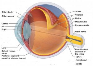
The wall of eyeball is made up of three layers
• Fibrous tunic:- it is the superficial layer of eyeball and consist of 2 parts- the anterior cornea and the posterior sclera. the cornea is a transparent coat that covers the iris and its curved shape helps focus light on the retina the cornea has 3 surfaces , the outer surface consist of non keratinized stratified squamous epithelium, the middle portion is of collagen fibers and fibro blasts while interior portion is simple squamous epithelium. The sclera is the white portion of the eye, it is a layer of dense connective tissue made up of collagen fibers and fibro-blasts, it covers the entire eye ball except the cornea , gives shape to the eyeball, makes it more rigid and also protects its inner parts and also works as site of attachment of extrinsic eye muscles. At the junction of cornea and sclera is an opening called as canal of Schlemm, aqueous humor drains in this opening.
• Vascular tunic – it is also known as Uvea, is the middle layer of the eyeball and consist of 3 parts
1. Choroid:- present on the posterior side of vascular tunic and is highly vascularized. It lines most of the internal surface of sclera. It provides nutrition to the part of retina, it also contain melanocytes because of which it is brown in color. Melanin also absorbs the stray light rays which prevents reflection and scattering within the eye, as the result the image appears sharp and clear.
2. Ciliary body:- in the anterior surface of this layer choroid becomes ciliary body, it also contains melanocytes , it also consist of ciliary processes and ciliary muscles. The ciliary processes are the fold in the internal surface and it secrets aqueous humor while ciliary muscles are smooth muscle whose contraction and relaxation which changes the position of lens.
3. Iris:- it is the colored portion of eye and has shape like a donut , it consist of melanocytes radial muscles and smooth muscles. The amount of melanin present in the iris is responsible for the color of the eye, blue color when melanin concentration is low, green when concentration is moderate and brown when high amount of melanin is present. The main function of iris is to regulate the size of pupil through the radial and circular muscles.
• Retina:- the inner layer of eyeball forms the posterior portion of the eye and visual pathway begins from retina. It consist 2 layers , the pigmented layer and neural layer; the pigmented layer is sheet of epithelium containing melanin and is present between the choroid and the neural layer, while the neural layer is the multilayered outgrowth of the brain and it processes the visual pathway.
Three types of layers of retinal neurons are also present here which are- photoreceptor layer, bipolar cell layer and ganglia cell layer. Two types of Photoreceptor are rods and cones , rods allow us to see in dim light while cones allow us to see in bright light.
Optic disc is the site where the optic nerve leaves the eyeball, the optic disc is also called as blind spot because of the absence of rods and cones in this region. Information flows from photoreceptors to the bipolar cells and then reaches the ganglia cells and through optic nerve reaches the brain.
MULTIPLE CHOICE QUESTIONS(MCQs)
1. the wall of eyeball is made up of how many layers?
A. 3 B. 4
C. 2 D. 5
2. Which of the following is the function of sclera?
A. gives the shape to eyeball B. makes it more rigid
C. protects inner parts D. all of the above
3. Out of the following options which one is NOT the accessory structure of eye?
A. Eyelids B. the lacrimal apparatus
C. intrinsic eye muscles D. eyebrows
4. Which of the following statement is NOT true?
a. cornea is highly vascularized structure
B. vascular tunic is also known as uvea
C. the junction of sclera and cornea forms scleral venous sinus
D. melanin prevents reflection and scattering of light within eye
5. In what condition the color of eye appears as blue?
A. When the concentration of melanin is high in iris
B. When the concentration of melanin is moderate
C. When the concentration of melanin is very low
D. none of the above
6. What causes the size of pupil to decrease during bright light
A. contraction of circular muscles of iris
B. contraction of radial muscles of iris
C. relaxation of circular muscle of iris
D. relaxation of radial muscle of iris
7. Which one is the only body part where the blood vessels can be viewed directly?
A. iris B. heart
C. retina D. choroid
8. Match the following:-
A. cornea 1. Rainbow, shaped like a donut
B. ciliary processes 2. Transparent coat that covers iris
C. iris 3. White portion of eye
D. sclera 4. Folds present in the internal
surface of ciliary body
9. Which of the following is the retinal neurons present in ganglia cell layer?
A. rods B. cones
C. amacrine cells D. none of the above
10. Which structure is also known as the “blind spot”?
A. optic disc B. macula lutea
C. cornea D. ciliary body
ANSWERS:-
1. 3
2. all of the above
3. intrinsic eye muscle
4. cornea is highly vascularized structure
5. When the melanin concentration is very low
6. contraction of circular muscle of iris
7. retina
8. A – 2 B – 4 C – 1 D – 3
9. none of the above
10. optic disc
Participate in Online FREE GPAT TEST: CLICK HERE
Participate in Online FREE Pharmacist TEST: CLICK HERE
Participate in Online FREE Drug Inspector TEST: CLICK HERE
REFRENCE: 1. Ross and Wilson-Anatomy and physiology in health and illness; 12th edition; page no.-: 196-199.
2. Gerard J. Tortora -Principles of anatomy and physiology; edition twelfth ; page no.-: 604-610.