AXIAL SKELETON: Hyoid bone, Vertebral column and Thorox and MCQs for NEET, GPAT
AXIAL SKELETON:
Hyoid Bone
It is a unique part of axial skeleton, as this bone does not articulate with any other bone. It is located in the anterior neck just between the mandible and larynx. The hyoid bone keeps the larynx open all the time; also the bone supports tongue and also serves as a site of attachment for tongue, neck and pharynx muscles. The bone consist of horizontal body and two projections: lesser horns and greater horns.
![]()
Vertebral Column
There are 26 bones in the vertebral column. It is also called as backbone, spine or spinal column. The vertebral column is divided in different regions, which are discussed below-
1.CERVICAL VERTEBRAE
These are the smallest vertebrae among all other, except from those who forms coccyx. Their vertebral arch is longer. all the cervical vertebrae have 3 foramina: one vertebral foramina and two transverse foramina. The first two cervical vertebrae are atypical(different from others).
The first cervical vertebrae(C1) is atlas, on which the skull rests. It is essentially a ring of bone with no distinct body or spinous process. It has two small transverse process. It also have two facets with articulates with the occipital bone, through condyloid joint and they allow nodding movement.
Just below the atlas, is the second cervical vertebrae(C2) known as axis. It has small body and a superior projection known as odontoid process. This process lies on the posterior foramen of atlas and is held in position by transverse ligaments. This joint allows side to side movement.
THORACIC VERTEBRAE
The 12 thoracic vertebrae are larger than the cervical vertebrae. It is because that this part of vertebral column has to support more body weight. The facets of bodies and transverse process of thoracic vertebrae articulates with the ribs, except of T11 and T12. These joints are called as vertebrocostal joints. Movements of thoracic region are limited due to the attachment of ribs to the sternum.
LUMBER REGION
The 5 lumber vertebrae are the largest and strongest of the vertebral column as it has to bear the weight of upper body. They have spinous process to which the muscles of lower neck attaches.
SACRUM VERTEBRAE
The sacrum is a triangular bone which Is formed from the union of 5 bones. It has concave anterior surface. The upper part called as base articulates with the L5. On each side, sacrum articulates with the ilium of hip bone to form sacroiliac joint; and at its lower end, it articulates with the coccyx. The vertebral foramina are present; also many foramina are present on each side which allows passage for nerves.
COCCYX VERTEBRAE
The coccyx is a triangular bone, formed by the fusion of 4 coccygeal vertebrae. On lateral surface, are the series of transverse processes in which the pair is the largest. At the upper end, the base of coccyx articulates with the apex of sacrum. In females, the coccyx points inferiorly to allow passage for baby during birth; while in male, the coccyx points anteriorly.

Above Figure is taken for Educational Purpose Only.
Features of the vertebral column:-
- a. Intervertebral discs:- The body of adjacent vertebrae are separated by the intervertebral discs; it consist of outer rim made f fibrocartilage and central core made of soft gelatinous material.
- b. Intervertebral foramina:- When the two adjacent vertebrae are viewed from side , a foramen formed is seen by the gap between the two vertebrae.
- c. Ligaments of vertebral column:- These ligaments holds the vertebral column together and also holds the intervertebral discs in proper position.
- d. Curves of the vertebral column:- The vertebral column has four curves:- 2 primary curves and 2 secondary curves. When the fetus in the uterus lies in the curled position, it shows primary curve. The secondary cervical curve develops when the child can hold up his head; and the secondary lumber curve develops when the child is able to stand.
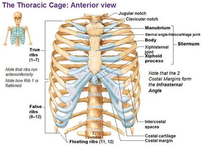
Thoracic Cage
The thoracic cage is the skeletal part of the thorax which consist of the ribs, sternum, their costal cartilages and the bodies of thoracic vertebrae. It protects the organs of thoracic and abdominal cavities; and also provides support to the bones of upper limb.
STERNUM
It is a flat bone which lies in the middle of the front of the chest. It is divided in 3 regions:-
- The manubrium is the uppermost part of the sternum and articulate with the clavicle to form sternoclavicular joint. It also articulates with the first two pairs of ribs
- The body is the middle portion of sternum. It provide attachment for the ribs
- The xiphoid process It is the inferior end of the sternum. It provides attachment to the diaphragm and anterior muscles of abdominal wall.
RIBS
The 12 pairs of ribs gives the structural support to the side of the thoracic cavity. The ribs increases in length till 7th pair; and then after decreases in length till 12th pair. Each ribs articulates posteriorly with its corresponding thoracic vertebrae.
The first seven pairs of ribs have direct anterior attachment with the sternum by the strip of hyaline cartilage known as costal cartilage. The ribs that have costal cartilages and attaches directly to the sternum are known as true ribs. The joints between the true ribs and the sternum are called as sterno-costal joints. The next 3 pairs (8, 9, 10th) are called as false ribs because their costal cartilages attaches indirectly to the sternum. The rest two pairs are called floating rib because their costal cartilage do not attach to the sternum at all.
Inflammation of any one or more costal cartilage is known as costochondritis. It causes local chest wall pain; and be resulted as a symptom for heart attack.
Multiple choice questions(MCQs)
1. Which is the only bone of axial skeleton which does not articulate with any other bone?
A. occipital bone B. mandible
C. hyoid bone D. none of the above
2. How many bones form the vertebral column?
A. 31 B. 26
C. 22 D. 30
3. Which type of joint lies between the atlas and the occipital bone?
A. saddle joint B. pivot joint
C. hinge joint D. condyloid joint
4. Which bone of vertebral column facilitates side to side movements?
A. C7 B. C5
C. C4 D. C2
5. Match the following:-
A. sacrum vertebrae 1. Articulates with the ribs
B. coccyx vertebrae 2.articulates with the ilium
C. lumber vertebrae 3. Bears upper body weight
D. thoracic vertebrae 4. The lateral surface has transverse process
6. Which of the following is the part of sternum?
a. body B. xiphoid process
C. manubrium D. all of the above
7. Which connective tissue provides the attachment of the true ribs with sternum?
A. hyaline cartilage B. areolar tissue
C. fibrocartilage D. elastic cartilage
8. Which of the following statement is true?
A. transverse process of Lumber attaches neck muscle
B. the joint between C1 and occipital bone allows nodding
C. all the thoracic vertebrae articulates with ribs
D. the base of sacrum articulates with ilium to form sacroiliac joint
9. Which of the following is known as floating ribs?
A. 7th pair B. 10th pair
C. 9TH pair D. none of the above
10. What is costochondritis?
A. inflammation of ribs
B. inflammation of manubrium
C. inflammation of costal cartilage
D. inflammation of xiphoid process
ANSWERS:-
1. hyoid bone
2. 26
3. condyloid joint
4. C2
5. A – 2 B – 4 C – 3 D – 1
6. all of the above
7. hyaline cartilage
8. the joint between C1 and occipital bone allows nodding
9. none of the above
10. inflammation of costal cartilage
Participate in Online FREE GPAT TEST: CLICK HERE
Participate in Online FREE Pharmacist TEST: CLICK HERE
Participate in Online FREE Drug Inspector TEST: CLICK HERE
REFRENCE: 1. Ross and Wilson-Anatomy and physiology in health and illness; 12th edition; page no.-:401-406.
2. Gerard J. Tortora -Principles of anatomy and physiology; edition twelfth ; page no.-: 216-226.