Nervous Tissue Anatomy and MCQs for NEET, GPAT, CSIR NET JRF, Staff Nurse Exam
Nervous tissue is of two types: neurons and neuralgia. Neurons are the working unit of the nervous system that generate nerve impulses. These neurons are supported by the connective tissue; collectively these tissue are known as neuralgia. Neuralgia is formed from different types of glial cells.
NEURONS:-
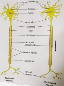
Each neuron consist of cell body and its processes: one axon and numerous dendrites. These neurons are also called as nerve cells. Bundle of the axons is known as nerve. These neurons need oxygen and glucose on regular basis to perform there work.
These neurons generate and transmit nerve impulses called as action potential. Some neurons generate the impulses while other neurons for the transmission of these signals ahead.
Nerve impulses are stimulated in to response to:
• Outside body-eg touch, light ect
• Inside the body- eg: change in concentration of CO2 in the blood can affect respiration.
The transmission of signal is sometime electrical and sometimes chemical; When the nerve impulses travel along axons, it is electrical signal; but when the signal flows from one nerve cell to the another, it is chemical because the nerves do not come in direct contact with each other.
Cell bodies
Nerve cells vary in shape and size and are very small to be seen from the naked eye. The cell bodies form the grey matter of the nervous system. This grey matter is found at the periphery(sides) of brain and in the center of spinal cord. Group of these cell bodies are called nuclei( in CNS) and ganglia(in PNS)
Axons
Axons and dendrites are the extension of the nerve cell and form the white matter of nervous system. Axons are found deep in the brain in tracts. These axons are referred as nerve or nerve fibers outside the brain and spinal cord. Each nerve cell has only 1 axon; which starts from the the axon hillock. They carry nerve impulses ahead of the nerve cell and are longer than dendrites. The membrane of the axon is called as axolemma and it surrounds the cytoplasm of the cell body. There are two types of neurons:-
- myelinated neuron: these are large axons; and those of peripheral nerves are surrounded by myelin sheath. It consist of a large number of schwann cells which lies along the length of the axon. each cell is covered by a multiple layered plasma membrane. Between the layers of plasma membrane is a fatty substance present known as myelin. Nodes of Ranvier are the tiny areas exposed between the adjacent schwann cell. These nodes carry out rapid transmission of impulses in myelinated neurons.
- Unmyelinated neuron: these are the postganglionic fibers; and small fibers of CNS. In this type of neuron, a number of axons are embedded in 1 Schwann cell. There is no exposed axolemma and the Schwann cells are in close association with each other. Transmission of impulses is slower than in myelinated neurons.

Dendrites
These are short processes that carry and receive the impulses from and towards the cell body. They have same structure as axons; but are smaller than axons. In motor neurons, dendrites form a part of synapses; and in sensory neurons, they acts as sensory receptors and responds to a particular stimulation.
Nerves
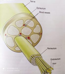
A nerve consist of a number of neurons which are collected into bundles. Each bundle has several coverings of connective tissue for protection. These are:-
• Endoneurium:- it is a delicate tissue which surrounds individual fiber
• Perineurium:- it is a smooth connective tissue which covers each bundle of fibers
• Epineurium:- fibrous tissue which surrounds and protects a number of bundle of nerve fibers.
Nerves are of 3 types:-
1. Sensory or afferent nerves- these carry info from the body to the spinal cord; and then to the brain or connector neuron or reflex arcs. These nerves have sensory receptors which respond to the changes inside and outside the body
2. Motor or efferent nerves- these originate from the brain, spinal cord and autonomic ganglia(ANS). They transmit impulses to the effector organ, muscles and glands. Two types of motor nerves are present: somatic nerves which are involved in voluntary control of skeletal muscle contraction; autonomic nerves which are involved in involuntary control.
3. Mixed nerves- These nerves contain both sensory as well as motor fibers. Like the spinal cord has sensory and motor fibers in different tracts; but as nerve leaves the cord, these fibers are enclosed in same connective sheath.

NEUROLGIA
The neurons of CNS are supported by glial cells. Unlike the nerve cells which cannot divide, the glial cells continue to replicate throughout the life. There are four types:-
- Astrocytes: These cells form the main supporting tissue in CNS. These cells are star shaped. Example is the blood-brain barrier; a selective barrier which protects the brain from the toxic substances and chemical variations of blood.
- Oligodendrocytes: These are smaller cells than astrocytes and are found in round nerve cell bodies of grey matter; where They provide support. These cells form and maintain myelin like Schwann cells in PNS
- Ependymal cells: They form the covering of the ventricles of brain and the central canal of spinal cord. These cells form choroid plexus that secrete cerebrospinal fluid.
- Microglia: They are least in number, and are formed from the monocytes that migrate from blood to the nervous system before birth.
Multiple choice questions(MCQs)
1. How many types of nervous tissue are present in the body?
A. 2 B. 3
C.1 D. 4
2. Which structure is known as the working unit of nervous system?
A. nerves B. neurons
C. cell body D. neuralgia
3. Which of the following is a part of neuron?
A. axon B. cell body
C. dendrite D. all of the above
4. Match the following-
A. astrocytes 1. Forms the choroid plexus of ventricles
B. microglia 2. These are star shaped cells
C. ependymal cells 3. Covers the cell body in grey matter
D. oligodendrocytes 4. Formed from monocytes
5. When is the transmission of signal electrical?
A. when signal travels along the axon
B. signal flows from one nerve cell to another
C. never
D. always
6. What forms the grey matter of nervous system?
A. axon B. cell body
C. neuralgia D. dendrites
7. Which of the following statement is NOT true?
A. nerves are bundles of axons
B. neuralgia is a connective tissue that protects neurons
C. motor nerves are of 3 types
D. axons begin at the axon hillock
8. Which structure of myelinated neurons carry out transmission of signals?
A. Schwann cell B. nodes of Ranvier
C. axolemma D. axon hillock
9. What is the role of dendrites in case of motor neurons?
A. acts as sensory organ B. forms synapse
c. works as receptors D. none of the above
10. Which layer of connective tissue of nerve covers the individual fiber?
A. perineurium B. pericardium
c. epineurium D. none of the above
ANSWERS:
1. 2
2. neurons
3. all of the above
4. A – 2 B – 4 C – 1 D – 3
5. when signal travel along the axons
6. cell body
7. motor nerves are of 3 types
8. nodes of Ranvier
9. forms synapse
10. none of the above
Participate in Online FREE GPAT TEST: CLICK HERE
Participate in Online FREE Pharmacist TEST: CLICK HERE
Participate in Online FREE Drug Inspector TEST: CLICK HERE
REFERENCE:
1. Ross and Wilson-Anatomy and physiology in health and illness; 12th edition; page no.-: 144-151
- Gerard J. Tortora -Principles of anatomy and physiology; edition twelfth ; page no.-: 416-421.